Science & A.I. · Neuroscience & medicine
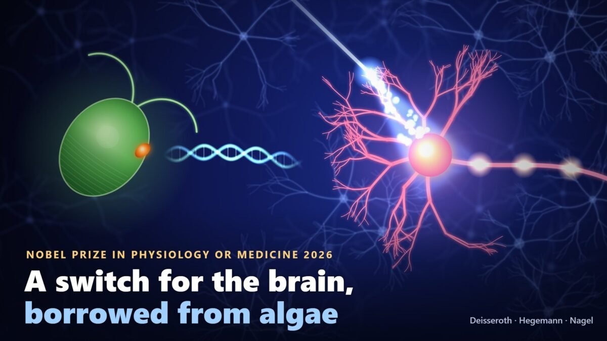
A green alga too small to see swims towards the light. Asking how it manages that turned out to be one of the most productive questions in modern biology, and on 5 October 2026 it earned three scientists the Nobel Prize in Physiology or Medicine.
The Nobel Assembly at Karolinska Institutet awarded the prize jointly to Karl Deisseroth of the Howard Hughes Medical Institute and Stanford University, Peter Hegemann of the Humboldt University of Berlin and Georg Nagel of the University of Würzburg, “for their discoveries concerning light-gated ion channels and optogenetics”. The three share 12 million Swedish kronor.
“Optogenetics provides opportunities for mapping the brain in a way that we could once only dream of,” said Per Svenningsson, chair of the Nobel Committee for Physiology or Medicine, in the official announcement.
The idea itself is simple enough to explain at a kitchen table. Take a gene from an alga that makes a light-sensitive protein, put it into a chosen type of nerve cell in an animal’s brain, and those cells, and only those, can now be switched on by a flash of blue light. For the first time, scientists could flip a specific group of neurons on or off, in a living animal, within a thousandth of a second, and watch what happened.
We wanted to see how far that has travelled. On 9 October 2026, PubMed, the US National Library of Medicine’s index of biomedical research, listed 15,726 papers mentioning optogenetics. The public trial registry ClinicalTrials.gov listed 15 studies in people. Every one of those 15 concerns the eye.
That gap is not a scandal. Optogenetics was built as a tool for discovery, and most of its medical value so far has come indirectly, by showing which brain circuits matter in disease. But it is the essential context for reading the celebrations. This article explains what the laureates found, how a question about pond algae became a switch for the brain, what the first patients have experienced, and what the method still cannot do.
What the 2026 Nobel was awarded for
The prize honours two linked achievements. Hegemann and Nagel discovered channelrhodopsins, proteins in a single-celled alga that act as both a light sensor and a gate in the cell’s outer membrane. Deisseroth showed that one of them could be moved into mammalian nerve cells and used as a switch, launching the method now known as optogenetics.
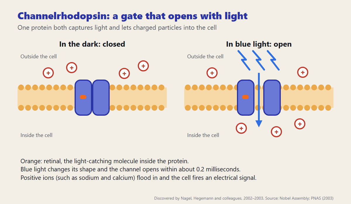
A nerve cell communicates with electricity. Charged particles called ions flow through gates in its membrane, and when enough flow in, the cell fires a brief electrical pulse that passes a signal to its neighbours. Channelrhodopsin is such a gate, but one that opens when light falls on it. Install it in a neuron, shine the right colour of light, and the neuron fires.
What makes this powerful is precision. Genetic tricks can restrict the gate to one type of neuron among many tangled together, and light can be switched in less than a millisecond. Researchers can therefore ask a question the brain sciences had struggled with for a century: not just which cells are active during a behaviour, but whether those cells cause it.
The problem: a brain you could watch but not steer
The human brain holds about 86 billion neurons, according to a careful 2009 count, each connected to thousands of others. Cells with completely different jobs sit side by side, and a single neuron can send its fibre to a distant region. That density defeated the tools neuroscience had for most of the twentieth century.
Scientists could record activity with electrodes or brain scans, but that showed only correlation: a region lights up during fear, which does not prove it produces fear. They could damage a region, or stimulate it with an electrode, but an electrode excites every cell near its tip, and a lesion destroys everything in its path. Drugs act too slowly and spread too widely to follow the millisecond rhythm of thought.
Francis Crick, who shared the 1962 Nobel for the structure of DNA and later turned to the brain, urged neuroscientists in a 1999 paper to tell molecular biologists what tools they needed. As the Nobel committee recounts, he imagined using light to control chosen nerve cells, an idea he admitted sounded far-fetched. The answer was already being worked out, in a lab that studied algae.
A question about pond algae
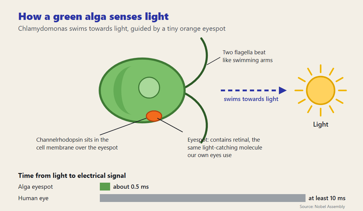
In the early 1990s, Peter Hegemann, then at the Max Planck Institute for Biochemistry in Martinsried near Munich, asked how Chlamydomonas, a single-celled green alga, could respond to light so quickly. Stir the algae into a dish and light one side, and the faint green colour drifts towards the light as millions of cells swim towards it. They sense it through an eyespot, a tiny orange patch containing retinal, the same light-catching molecule our eyes use.
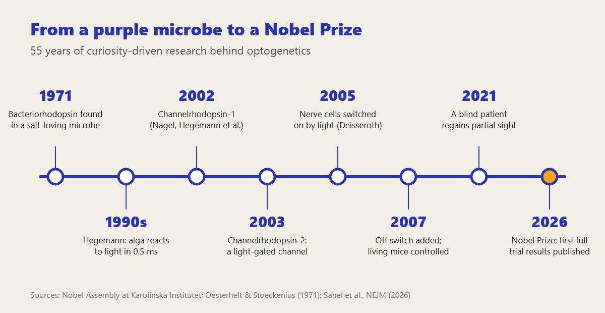
Using minuscule electrodes, Hegemann found that an electrical signal arose in the alga about half a millisecond after light reached the eyespot. In the human eye, light sets off a chain of chemical steps before an ion channel finally opens, and the process takes at least 10 milliseconds. The alga was more than twenty times faster.
Hegemann proposed the simplest explanation: in the alga, a single protein must both capture the light and form the channel. Colleagues were sceptical. Many kinds of ion channel were known, but none that responded to light on its own. For years the protein resisted isolation, falling apart whenever it was removed from the algal eyespot.
Frog eggs and a light-gated channel
The breakthrough came from genetics. Around the turn of the millennium, Japanese researchers published sequences for thousands of Chlamydomonas genes, and Hegemann’s group spotted two that resembled known light-sensing proteins. Hegemann sent them to Georg Nagel at the Max Planck Institute for Biophysics in Frankfurt, who specialised in studying membrane proteins by making frog eggs produce them.
Nagel injected each gene into eggs of the African clawed frog, which then built the algal protein into their outer membrane. When he shone light on them, current flowed. In 2002, in Science, the team described channelrhodopsin-1 as a light-gated channel for protons. In 2003, in the Proceedings of the National Academy of Sciences, they showed that channelrhodopsin-2 lets positively charged ions such as sodium and calcium flood in, and concluded that it could be used to excite cells “simply by illumination”. According to the Nobel committee, the channel opened within 0.2 milliseconds of a light pulse.
Science rarely moves in one lane. In June 2002 a team led by John Spudich at the University of Texas independently reported that the same two algal proteins control how Chlamydomonas swims towards light. The biophysicist Ernst Bamberg, Nagel’s colleague in Frankfurt, was a co-author on the key channelrhodopsin papers. The Nobel citation names three people, but the discovery grew out of a community of labs.
A thread back to a purple microbe
The algal protein had an ancestor in the literature. In 1971 Dieter Oesterhelt and Walther Stoeckenius described a rhodopsin-like protein in the purple membrane of a salt-loving microbe, then called Halobacterium halobium. The protein, later named bacteriorhodopsin, was soon shown to use light to pump charged particles across the membrane, proof that a single protein holding retinal could turn light into an electrical effect.
That work founded the study of microbial rhodopsins, the family to which channelrhodopsin belongs. In 2021 Oesterhelt, Hegemann and Deisseroth shared the Albert Lasker Basic Medical Research Award, one of America’s most prestigious biomedical prizes, for discovering these proteins and turning them into optogenetics. The thread from a purple microbe in 1971 to a Nobel in 2026 runs for 55 years, almost all of it curiosity-driven basic research.
Stanford: from algae to nerve cells
Karl Deisseroth came to the problem from the clinic. Training in medicine at Stanford in the 1990s, he spent time on a psychiatric ward and was struck by how little doctors could do for patients with severe depression or schizophrenia, and how often the treatments carried heavy side effects. He took a doctorate in neuroscience alongside his medical degree and concluded that understanding such illnesses required a way to control specific cells in a living brain.
When his new lab heard about channelrhodopsin-2, Deisseroth wrote to Nagel asking for the gene. His team packaged it into a modified virus that carries genes into cells, delivered it to rat neurons growing in a dish, and shone blue light on them. The neurons fired. In 2005 the group reported in Nature Neuroscience reliable control of nerve cell firing on a millisecond timescale. The paper’s first two authors were Ed Boyden and Feng Zhang; Zhang later became one of the pioneers of CRISPR gene editing.
The result was decisive because nothing about it was exotic. The algal protein worked in mammalian neurons without extra ingredients, because animal cells already contain the retinal it needs. Any lab that could deliver a gene could now control neurons with light.
Switching neurons off as well as on
A switch that only turns things on answers half the question. In 2007 Deisseroth’s group, with collaborators including Nagel and Bamberg, added an off switch: halorhodopsin, a light-driven pump from the archaeon Natronomonas pharaonis that pushes negatively charged chloride ions into a cell and silences it. Activated by yellow light rather than blue, it could be used in the same cells as channelrhodopsin-2, so researchers could excite or silence neurons on command.
By then the method had a name. In 2006 it was christened optogenetics, joining optics, the control of light, with genetics, the targeting of specific cells. Since then dozens of variants have been found or engineered, responding to different colours or to much dimmer light.
Into the living brain
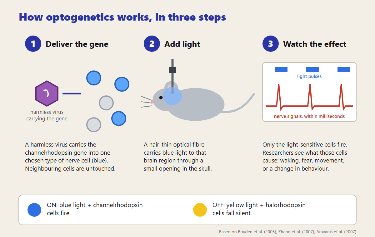
In 2007 Deisseroth’s team took the switch into living animals. They delivered channelrhodopsin-2 to cells in the motor cortex of mice, slid a hair-thin optical fibre through a small hole in the skull, and moved the animals’ whiskers by switching the light on and off.
The same year, working with Luis de Lecea’s group at Stanford, they put the switch into hypocretin neurons, a small population in the hypothalamus whose loss has been linked to narcolepsy. Flashing light into those cells woke sleeping mice faster, but only when the pulses came 5 to 30 times a second; a slow flash once a second did nothing. It was among the first demonstrations that a defined group of cells, firing in a particular rhythm, causes a specific mammalian behaviour.
What light-switched neurons have revealed
Some of the most striking results concern memory. In 2012 Susumu Tonegawa’s group at the Massachusetts Institute of Technology labelled the neurons in a mouse’s hippocampus that were active while it learned to fear a particular box. Later, in a different and safe box, switching those cells on with light made the mouse freeze in fear, though nothing frightening was happening. A memory, or at least its trace in a small set of cells, could be recalled on demand. The finding sharpened a question explored in our article on why science still cannot explain consciousness: knowing which cells hold a memory is not the same as knowing what it is like to remember.
Other studies have taken apart behaviours piece by piece. A 2018 study from Catherine Dulac’s lab at Harvard found that separate groups of neurons in a mouse’s hypothalamus control different parts of parenting, such as gathering pups into the nest and grooming them. Light could tune one component without touching the others.
The method has also crossed the boundary between brain and body. In 2023 Deisseroth’s team built a light-driven pacemaker that could race a mouse’s heart to as fast as 900 beats a minute. A faster heartbeat alone made the animals more anxious, but only in risky surroundings, and the effect ran through a brain region called the posterior insula. In 2022 a team at Duke University used an optical fibre in the gut to show that mice prefer real sugar to sweetener because of specialised sensor cells in the intestine that signal the brain.
Optogenetics has helped medicine indirectly, too. Deep brain stimulation, in which implanted electrodes deliver pulses to ease Parkinson’s disease, worked for years without anyone being sure which cells it acted on. A 2009 optogenetic study in rodents suggested that its benefit in one key target, the subthalamic nucleus, comes from stimulating incoming nerve fibres rather than the local cells, an insight into how an established treatment actually works.
Not the only route to the idea
A Nobel Prize can go to at most three people, and optogenetics had more pioneers than that. In January 2002 Gero Miesenböck, then at Memorial Sloan Kettering in New York, made neurons light-sensitive by giving them three fruit-fly genes from the eye, a system he called chARGe. In 2005, now at Yale, he and Susana Lima used light to trigger escape jumps and changes in movement in fruit flies by activating chosen neurons.
Miesenböck’s system was slower and more complicated than a single algal protein, which is why channelrhodopsin became the standard tool. But the principle he demonstrated was the same. In 2013 the Brain Prize, a major European neuroscience award, was shared by six people for optogenetics: Bamberg, Boyden, Deisseroth, Hegemann, Miesenböck and Nagel. The difference between six names and three is a reminder that prizes simplify how science actually happens.
From mice to people: the first patients
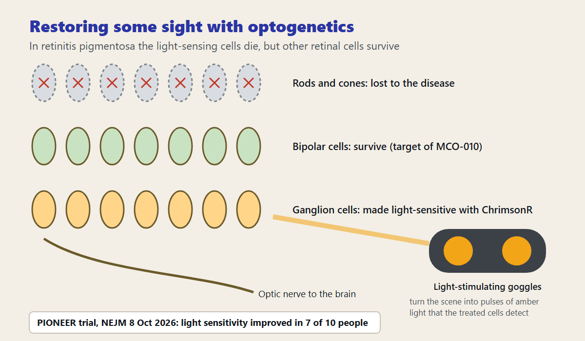
The first medical use of optogenetics is in the eye. Retinitis pigmentosa is a group of inherited diseases, linked to well over 100 genes, in which the light-sensing rods and cones of the retina die. People typically lose night vision in adolescence, side vision in young adulthood and central vision later in life. But other cells in the retina, including the ganglion cells that carry signals to the brain, often survive, and optogenetics can make them sensitive to light.
In 2021 José-Alain Sahel and colleagues reported in Nature Medicine on a patient blinded by the disease who received an injection into one eye of a harmless virus carrying ChrimsonR, a channelrhodopsin that responds to amber light. Wearing goggles that convert the scene into pulses of that light, the patient could perceive, locate, count and touch objects on a table using the treated eye. Before the treatment, or without the goggles, none of the objects could be seen. Recordings from the scalp showed activity over the visual cortex during the task.
On 8 October 2026, three days after the prize, the full results of that first safety trial appeared in the New England Journal of Medicine. Ten people with advanced retinitis pigmentosa received the injection. Nine had eye-related side effects, 34 events in all, most mild or moderate. One was severe: a blockage of the main artery of the retina immediately after the injection, which cleared within minutes with treatment. Light sensitivity improved in seven of the ten, by factors from 2 to 62, and in six the improvement met the threshold the researchers considered meaningful. The authors call the treatment safe within the limits of a small study and say more research is needed.
A second approach is closer to the market. Nanoscope Therapeutics’ MCO-010, which delivers a gene for an engineered light-sensitive protein to the retina’s bipolar cells, was tested in a 27-patient phase 2 trial called RESTORE; the company reports improved visual acuity at 52 and 76 weeks. In September 2026 the US Food and Drug Administration accepted the company’s application for approval, under the proposed name Mogenry, and the company expects a decision in the first half of 2027. An independent review by the Institute for Clinical and Economic Review in May 2025 found the evidence adequate to show a net health benefit but flagged uncertainty about how well and how long it works.
Why the eye goes first, and what comes next
The eye is the natural starting point. It is transparent, so light reaches the treated cells without implants. It is small and enclosed, so a single injection can reach its target. And people who have lost their sight have a clear, measurable outcome to gain. The brain offers none of those advantages: light has to be piped in through implanted fibres or devices, and light scatters quickly in tissue.
Hearing may be next. Today’s cochlear implants stimulate the auditory nerve with electricity, which spreads and blurs the sound. The Nobel committee notes hopes that light-sensitive auditory neurons could be stimulated more precisely, and a team at the University Medical Center Göttingen has been developing an optical cochlear implant with the aim of human trials, though none has yet been reported.
For the brain itself, a 2025 roadmap in Nature Neuroscience, written by an international group of neuroscientists and ethicists, argued that the main medical payoff will often be indirect: optogenetic experiments reveal which cells and circuits drive a disease, and that knowledge is then turned into other kinds of treatment. The authors also warned that applying optogenetics directly in the human brain raises ethical questions that need careful consideration before it spreads beyond blindness.
What optogenetics cannot do yet
Every optogenetic experiment requires two interventions: a gene delivered into specific cells, usually by a modified virus, and light delivered to those cells. In people, the first is a form of gene therapy, with the risks of immune reactions and of the gene reaching unintended cells. The second, outside the eye, means surgery to implant a light source, with the risk of heat and tissue damage.
The method is also artificial in a way that matters for interpretation. Light drives large groups of cells to fire in unison, which is not how the brain normally works, and a behaviour produced by switching cells on shows what those cells can do, not necessarily what they usually do. Researchers increasingly combine optogenetics with recordings of natural activity to guard against over-interpretation.
Most of what has been learned comes from mice, flies and worms. Human brains are larger, and circuits found in rodents do not always map neatly onto people. No optogenetic treatment has yet been approved anywhere, and the first decisions, for the eye, are not expected before 2027.
Who the laureates are
Karl Deisseroth, born in 1971, earned his PhD in 1998 and his medical degree in 2000 at Stanford University, where he is the D.H. Chen Professor of Bioengineering and of Psychiatry and Behavioral Sciences, and a Howard Hughes Medical Institute investigator. He trained as a psychiatrist.
Peter Hegemann, born in 1954, took his PhD in 1984 at the Max Planck Institute for Biochemistry in Martinsried, where he made his prize-winning discoveries, and is now Hertie Senior Professor of Neuroscience at the Humboldt University of Berlin. Georg Nagel, born in 1953, took his PhD in 1988 at the University of Frankfurt, did the prize-winning work at the Max Planck Institute for Biophysics in Frankfurt, and is professor of molecular plant physiology at the University of Würzburg.
The prize continues a pattern visible in last year’s award, explored in our article on the 2025 Nobel Prize for the immune system’s peacekeepers: work that began with a question nobody thought practical becomes, decades later, the foundation of a field.
Why this prize matters beyond the laboratory
Nobody studying how an alga swims towards light was trying to treat blindness or map memory. Hegemann wanted to understand a fast electrical response in a single cell; Nagel was expert in getting membrane proteins to work in frog eggs; Deisseroth, a psychiatrist, was looking for a better way to study the brains of his patients. The tool emerged where those interests met.
That is a practical lesson about how discoveries happen, and it applies well beyond neuroscience. The gene-editing tools that now rewrite our DNA began with curiosity about how bacteria fight off viruses; the light switch for neurons began with an alga swimming towards the light. Both were found by people pursuing curiosity, often with public funding, long before anyone knew what they would be good for.
It is also a lesson in patience. Fifty-five years separate bacteriorhodopsin from this prize, and twenty-one years separate the first light-switched neurons from the first full trial results in patients. For the families waiting on treatments for blindness, Parkinson’s disease or depression, optogenetics is not yet a cure. It is something rarer and more durable: a way of asking the brain direct questions and getting answers.
Frequently asked questions
Who won the 2026 Nobel Prize in Medicine?
Karl Deisseroth of the Howard Hughes Medical Institute and Stanford University, Peter Hegemann of the Humboldt University of Berlin and Georg Nagel of the University of Würzburg. The Nobel Assembly at Karolinska Institutet announced the prize on 5 October 2026, and the laureates share 12 million Swedish kronor.
What was the prize awarded for?
The official citation is “for their discoveries concerning light-gated ion channels and optogenetics”. Hegemann and Nagel discovered channelrhodopsins in a green alga, and Deisseroth turned channelrhodopsin-2 into a light-controlled switch for nerve cells.
What is optogenetics, in simple terms?
It is a way of controlling chosen nerve cells with light. A gene for a light-sensitive protein, often taken from algae or microbes, is delivered into a specific type of cell. When light of the right colour reaches those cells, they switch on or off within milliseconds, letting researchers test what the cells actually do.
What is channelrhodopsin?
A protein found in the eyespot of the single-celled alga Chlamydomonas. It sits in the cell’s membrane, captures light with a molecule called retinal and opens a channel that lets charged particles flow in, producing an electrical signal. Channelrhodopsin-2 opens within a fraction of a millisecond.
Is optogenetics used to treat people?
Only experimentally, and so far only in the eye. In 2021 a blind patient with retinitis pigmentosa regained partial vision with an optogenetic gene therapy and special goggles, and a 10-person trial published in October 2026 found improved light sensitivity in seven participants. No optogenetic treatment has yet been approved, though the FDA is reviewing one for retinitis pigmentosa and its developer expects a decision in 2027.
Could optogenetics treat depression or Parkinson’s disease?
Not directly at present. In animals it has revealed circuits involved in mood, fear, reward and movement, and it has helped explain how deep brain stimulation eases Parkinson’s disease. Using it inside the human brain would require gene therapy plus an implanted light source, and experts have urged careful ethical scrutiny before that happens.
Why did the discovery come from algae?
Because algae need to sense light quickly to swim towards it, and evolution gave Chlamydomonas a protein that captures light and opens an ion channel in one step. Peter Hegemann noticed in the early 1990s that the alga responded to light far faster than the human eye, and suspected exactly such a protein.
Why were other optogenetics pioneers not included?
A Nobel Prize can be shared by at most three people. Other researchers made important contributions, including Gero Miesenböck, who made neurons light-sensitive in 2002 with fruit-fly genes, and Ed Boyden and Ernst Bamberg, co-authors of key papers. In 2013 the Brain Prize was shared by six optogenetics pioneers.
Is optogenetics safe?
In animal research it is widely used, but in people it combines the risks of gene therapy and light delivery. In the October 2026 eye trial, nine of ten participants had eye-related side effects, mostly mild or moderate, and one had a brief blockage of the retinal artery after the injection that resolved within minutes. Larger trials are needed.
How is optogenetics different from deep brain stimulation?
Deep brain stimulation uses an electrode that excites every cell near its tip. Optogenetics acts only on cells that carry the light-sensitive gene, so it can target one cell type among many intermingled ones, and it can switch cells off as well as on. That precision is why it is so valuable for research.
Further reading on Web News For Us
Sources
Nobel Prize documentation:
- The Nobel Prize in Physiology or Medicine 2026 — press release (Nobel Assembly at Karolinska Institutet, 5 October 2026)
- NobelPrize.org — Popular science background: a light-sensitive algal protein energised neuroscience
- NobelPrize.org — Scientific background: Optogenetics, discovery of a neuronal switch
Primary peer-reviewed research:
- Nagel et al. — Channelrhodopsin-1: a light-gated proton channel in green algae, Science 296, 2395 (2002)
- Nagel et al. — Channelrhodopsin-2, a directly light-gated cation-selective membrane channel, PNAS 100, 13940 (2003)
- Boyden, Zhang, Bamberg, Nagel & Deisseroth — Millisecond-timescale, genetically targeted optical control of neural activity, Nature Neuroscience 8, 1263 (2005)
- Zhang et al. — Multimodal fast optical interrogation of neural circuitry, Nature 446, 633 (2007)
- Deisseroth et al. — Next-generation optical technologies for illuminating genetically targeted brain circuits, Journal of Neuroscience 26, 10380 (2006)
- Adamantidis et al. — Neural substrates of awakening probed with optogenetic control of hypocretin neurons, Nature 450, 420 (2007)
- Oesterhelt & Stoeckenius — Rhodopsin-like protein from the purple membrane of Halobacterium halobium, Nature New Biology 233, 149 (1971)
- Sineshchekov, Jung & Spudich — Two rhodopsins mediate phototaxis to low- and high-intensity light in Chlamydomonas reinhardtii, PNAS 99, 8689 (2002)
- Zemelman, Lee, Ng & Miesenböck — Selective photostimulation of genetically chARGed neurons, Neuron 33, 15 (2002)
- Lima & Miesenböck — Remote control of behavior through genetically targeted photostimulation of neurons, Cell 121, 141 (2005)
- Crick — The impact of molecular biology on neuroscience, Philosophical Transactions of the Royal Society B 354, 2021 (1999)
- Azevedo et al. — Equal numbers of neuronal and nonneuronal cells make the human brain an isometrically scaled-up primate brain, Journal of Comparative Neurology 513, 532 (2009)
- Liu et al. — Optogenetic stimulation of a hippocampal engram activates fear memory recall, Nature 484, 381 (2012)
- Kohl et al. — Functional circuit architecture underlying parental behaviour, Nature 556, 326 (2018)
- Hsueh et al. — Cardiogenic control of affective behavioural state, Nature 615, 292 (2023)
- Buchanan et al. — The preference for sugar over sweetener depends on a gut sensor cell, Nature Neuroscience 25, 191 (2022)
- Gradinaru et al. — Optical deconstruction of parkinsonian neural circuitry, Science 324, 354 (2009)
- Hartong, Berson & Dryja — Retinitis pigmentosa, The Lancet 368, 1795 (2006)
Clinical and regulatory:
- Sahel et al. — Partial recovery of visual function in a blind patient after optogenetic therapy, Nature Medicine 27, 1223 (2021)
- Sahel et al. — Optogenetic therapy for restoring aspects of visual function, New England Journal of Medicine 395, 1399 (2026)
- Lüscher et al. — Roadmap for direct and indirect translation of optogenetics into discoveries and therapies for humans, Nature Neuroscience 28, 2415 (2025)
- ClinicalTrials.gov — registered studies mentioning optogenetics (queried 9 October 2026)
- Foundation Fighting Blindness — Nanoscope’s investigational RP treatment Mogenry advances toward FDA approval (September 2026)
- Institute for Clinical and Economic Review — final evidence report on sonpiretigene isteparvovec for retinitis pigmentosa (15 May 2025)
Other recognition of optogenetics:
- Max Planck Society — Lasker Award 2021 for the pioneers of optogenetics (Oesterhelt, Hegemann, Deisseroth)
- The Brain Prize — Optogenetics, 2013 (Bamberg, Boyden, Deisseroth, Hegemann, Miesenböck, Nagel)
Baryon. (2026, October 9). 2026 Nobel Prize in Medicine: How a Light-Seeking Alga Gave Science a Switch for the Brain. Web News For Us. https://webnewsforus.com/2026-nobel-prize-in-medicine-optogenetics/
Baryon. “2026 Nobel Prize in Medicine: How a Light-Seeking Alga Gave Science a Switch for the Brain.” Web News For Us, 9 October 2026, https://webnewsforus.com/2026-nobel-prize-in-medicine-optogenetics/. Accessed 9 October 2026.